How scan detect cancer in pets. Cancer is a word that no pet owner wants to hear, but unfortunately, it is a common and serious health problem for many dogs and cats.. Cancer is the uncontrolled and abnormal growth of cells or tissues in the body, and it can affect any organ or system. Some cancers are benign, meaning they do not spread to other parts of the body, while others are malignant, meaning they can invade and damage nearby tissues or organs, or metastasize to distant sites.
The good news is that there are many ways to diagnose and treat cancer in pets, and some of them are similar to those used in humans. One of the most important tools for detecting and evaluating cancer in pets is a scan, which is an imaging test that creates pictures of the inside of the body. Scans can help reveal the presence, location, size, shape, and activity of tumors, as well as their effects on normal tissues and organs. Scans can also help monitor the response to treatment and check for recurrence of cancer.
There are different types of scans that can be used for pets with cancer, and each one has its own advantages and limitations. In this article, we will discuss some of the most common scans for pets with cancer, how they work, what they can show, and what to expect before, during, and after the procedure.
X-rays
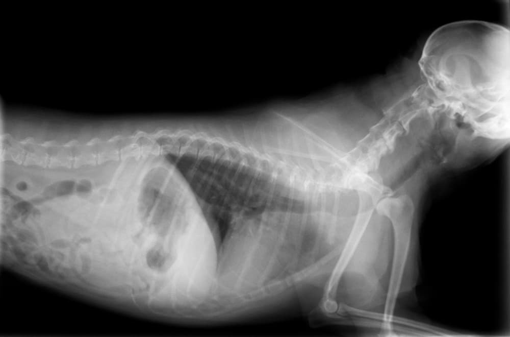
X-rays are the oldest and most widely used form of imaging for pets with cancer. X-rays use a beam of high-energy radiation that passes through the body and creates an image on a film or a digital screen. X-rays can show the shape and size of bones and organs, as well as the presence of foreign objects, fractures, infections, or tumors. X-rays are quick, painless, and relatively inexpensive, but they have some limitations. X-rays cannot show the difference between normal and abnormal tissues, such as cancer cells, and they cannot show the function or activity of organs or tumors. X-rays also expose the pet and the staff to radiation, which can be harmful in high doses or repeated exposures.
X-rays are often the first step in diagnosing cancer in pets, especially for bone tumors, lung tumors, or tumors that cause swelling or deformity of the body. X-rays can also be used to check for the spread of cancer to other parts of the body, such as the chest or the abdomen. X-rays can be performed in most veterinary clinics or hospitals, and they usually do not require any special preparation or sedation for the pet. However, some pets may need to be restrained or positioned in a certain way to get a clear image, which can cause some stress or discomfort. The pet owner may be asked to wait outside the room or wear a protective apron or shield to avoid exposure to radiation.
Ultrasound
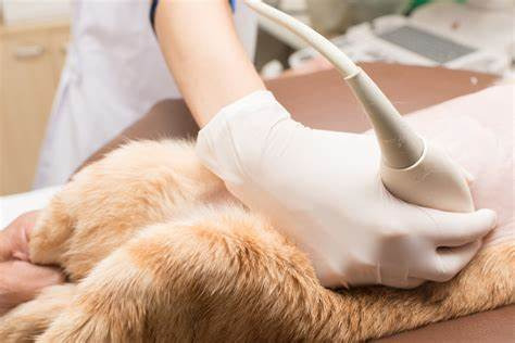
Ultrasound is another common and useful imaging test for pets with cancer. Ultrasound uses sound waves that bounce off the tissues and organs in the body and create an image on a monitor. Ultrasound can show the shape, size, texture, and blood flow of organs and tumors, as well as their relation to other structures in the body. Ultrasound can also help guide the placement of a needle or a probe for biopsy or treatment. Ultrasound is safe, painless, and non-invasive, but it also has some limitations. Ultrasound cannot penetrate bones or gas-filled organs, such as the lungs or the intestines, and it cannot show the function or activity of organs or tumors. Ultrasound also depends on the skill and experience of the operator, and it may not be available in all veterinary clinics or hospitals.
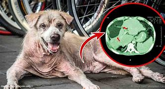
Ultrasound is often used to diagnose and evaluate cancer in pets, especially for abdominal tumors, such as liver, spleen, kidney, or adrenal tumors, or for tumors that involve the blood vessels, such as hemangiosarcoma. Ultrasound can also be used to check for the spread of cancer to the lymph nodes, the chest, or the heart. Ultrasound can be performed in most veterinary clinics or hospitals, and it usually does not require any special preparation or sedation for the pet. However, some pets may need to have their fur shaved or clipped in the area to be scanned, and some pets may need to be restrained or positioned in a certain way to get a clear image, which can cause some stress or discomfort. The pet owner may be allowed to stay with the pet during the procedure, or may be asked to wait outside the room.
Computed Tomography (CT) Scan

Computed tomography (CT) scan is a more advanced and sophisticated form of imaging for pets with cancer. CT scan uses a series of x-rays that rotate around the body and create a cross-sectional image of the tissues and organs. CT scan can show the shape, size, density, and location of organs and tumors, as well as their effects on normal tissues and organs. CT scan can also show the function or activity of organs or tumors by using a contrast agent, which is a substance that is injected into the blood or swallowed by the pet and makes the organs or tumors appear brighter or darker on the image. CT scan is more accurate, detailed, and comprehensive than x-rays or ultrasound, but it also has some drawbacks. CT scan is more expensive, time-consuming, and complex than x-rays or ultrasound, and it exposes the pet and the staff to more radiation. CT scan also requires the pet to be completely still and positioned in a certain way, which usually means the pet has to be sedated or anesthetized for the procedure.
CT scan is often used to diagnose and evaluate cancer in pets, especially for tumors that involve the head, neck, chest, spine, or limbs, such as brain, nasal, thyroid, lung, bone, or soft tissue tumors. CT scan can also be used to check for the spread of cancer to other parts of the body, such as the lungs, liver, or lymph nodes. CT scan can also be used to plan and monitor the treatment of cancer, such as surgery, radiation therapy, or chemotherapy. CT scan can be performed in some veterinary clinics or hospitals, or in specialized imaging centers. CT scan requires some special preparation and sedation or anesthesia for the pet. The pet owner may be asked to fast the pet for several hours before the procedure, and to remove any metal objects, such as collars, tags, or jewelry, from the pet. The pet owner may also be asked to sign a consent form and to leave the pet at the clinic or hospital for several hours or overnight.
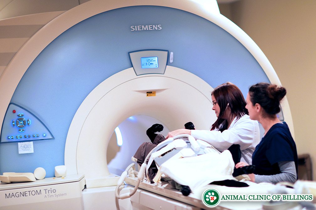
Magnetic Resonance Imaging (MRI) Scan
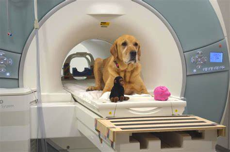
Magnetic resonance imaging (MRI) scan is another advanced and sophisticated form of imaging for pets with cancer. MRI scan uses a strong magnetic field and radio waves that interact with the water molecules in the body and create an image on a monitor. MRI scan can show the shape, size, structure, and composition of organs and tumors, as well as their effects on normal tissues and organs. MRI scan can also show the function or activity of organs or tumors by using a contrast agent, which is a substance that is injected into the blood or swallowed by the pet and makes the organs or tumors appear brighter or darker on the image. MRI scan is more accurate, detailed, and comprehensive than x-rays, ultrasound, or CT scan, but it also has some drawbacks. MRI scan is more expensive, time-consuming, and complex than x-rays, ultrasound, or CT scan, and it does not use radiation. However, MRI scan requires the pet to be completely still and positioned in a certain way, which usually means the pet has to be sedated or anesthetized for the procedure. MRI scan also requires the pet to be placed inside a narrow and noisy tube, which can cause some stress or discomfort. MRI scan also cannot be performed on pets that have metal implants, such as pacemakers, pins, or plates, as they can interfere with the magnetic field and cause damage or injury.
MRI scan is often used to diagnose and evaluate cancer in pets, especially for tumors that involve the brain, spinal cord, or nerves, such as brain, spinal, or nerve sheath tumors. MRI scan can also be used to check for the spread of cancer to other parts of the body, such as the brain, spinal cord, or nerves. MRI scan can also be used to plan and monitor the treatment of cancer, such as surgery, radiation therapy, or chemotherapy. MRI scan can be performed in some veterinary clinics or hospitals, or in specialized imaging centers. MRI scan requires some special preparation and sedation or anesthesia for the pet. The pet owner may be asked to fast the pet for several hours before the procedure, and to remove any metal objects, such as collars, tags, or jewelry, from the pet. The pet owner may also be asked to sign a consent form and to leave the pet at the clinic or hospital for several hours or overnight.
Positron Emission Tomography (PET) Scan
Positron emission tomography (PET) scan is a newer and more innovative form of imaging for pets with cancer. PET scan uses a radioactive substance called a tracer that is injected into the blood and accumulates in the areas of the body that have higher metabolic or biochemical activity, such as cancer cells. PET scan can show the presence, location, and activity of cancer cells, as well as their effects on normal tissues and organs. PET scan can also show the function or activity of organs or tumors by using different types of tracers, such as glucose, oxygen, or amino acids. PET scan is more sensitive, specific, and informative than x-rays, ultrasound, CT scan, or MRI scan, but it also has some drawbacks. PET scan is more expensive, time-consuming, and complex than x-rays, ultrasound, CT scan, or MRI scan, and it exposes the pet and the staff to radiation. PET scan also requires the pet to be sedated or anesthetized for the procedure, and it may cause some side effects, such as allergic reactions, nausea, or vomiting.
PET scan is often used to diagnose and evaluate cancer in pets, especially for tumors that are difficult to detect or measure by other imaging tests, such as lymphoma, melanoma, or mast cell tumors. PET scan can also be used to check for the spread of cancer to other parts of the body, such as the lungs, liver, bones, or lymph nodes. PET scan can also be used to plan and monitor the treatment of cancer, such as surgery, radiation therapy, or chemotherapy. PET scan can be performed in some veterinary clinics or hospitals, or in specialized imaging centers. PET scan requires some special preparation and sedation or anesthesia for the pet. The pet owner may be asked to fast the pet for several hours before the procedure, and to remove any metal objects, such as collars, tags, or jewelry, from the pet. The pet owner may also be asked to sign a consent form and to leave the pet at the clinic or hospital for several hours or overnight.
RELATED
How can pet allergies go away?
Conclusion
Cancer is a common and serious health problem for many pets, but there are many ways to diagnose and treat it, and some of them are similar to those used in humans. One of the most important tools for detecting and evaluating cancer in pets is a scan, which is an imaging test that creates pictures of the inside of the body. Scans can help reveal the presence, location, size, shape, and activity of tumors, as well as their effects on normal tissues and organs. Scans can also help monitor the response to treatment and check for recurrence of cancer.
There are different types of scans that can be used for pets with cancer, and each one has its own advantages and limitations. The most common scans for pets with cancer are x-rays, ultrasound, CT scan, MRI scan, and PET scan. Each scan works in a different way, and shows different aspects of the organs and tumors. Each scan also has different requirements, costs, and risks for the pet and the staff. The choice of the scan depends on the type, location, and stage of the cancer, as well as the availability and preference of the veterinarian and the pet owner.
Scans are not the only way to diagnose and treat cancer in pets, and they are not always accurate or conclusive. Scans may need to be combined with other tests, such as blood tests, urine tests, biopsies, or cytologies, to confirm the diagnosis and determine the best course of treatment. Scans may also need to be repeated or followed up with other scans, to monitor the progress or changes of the cancer. Scans are not a substitute for regular veterinary check-ups and preventive care, which can help detect and prevent cancer in pets.
If your pet has been diagnosed with cancer, or if you suspect that your pet may have cancer, you should consult with your veterinarian about the best options for your pet. Your veterinarian can explain the benefits and risks of each scan, and help you decide which one is most suitable for your pet. Your veterinarian can also provide you with more information and resources about cancer in pets, and support you and your pet throughout the process.
FAQs
- Q: How can I prepare my pet for a scan?
- A: The preparation for a scan depends on the type of scan and the condition of your pet. In general, you may be asked to fast your pet for several hours before the scan, and to remove any metal objects, such as collars, tags, or jewelry, from your pet. You may also be asked to sign a consent form and to leave your pet at the clinic or hospital for several hours or overnight. Your veterinarian will give you more specific instructions and advice on how to prepare your pet for a scan.
- Q: How can I comfort my pet during and after a scan?
- A: The comfort of your pet during and after a scan depends on the type of scan and the level of sedation or anesthesia required. In general, you may be allowed to stay with your pet during the scan, or you may be asked to wait outside the room. You may also be able to visit your pet after the scan, or you may have to wait until your pet is fully recovered and ready to go home. Your veterinarian will give you more specific instructions and advice on how to comfort your pet during and after a scan.
- Q: How can I interpret the results of a scan?
- A: The interpretation of the results of a scan depends on the type of scan and the expertise of the veterinarian or the radiologist. In general, you will receive a report that describes the findings and the impressions of the scan, and you will have a consultation with your veterinarian to discuss the results and the implications for your pet. Your veterinarian will explain the meaning and the significance of the scan, and answer any questions or concerns you may have.
- Q: How much does a scan cost for my pet?
- A: The cost of a scan for your pet depends on the type of scan and the location and the facility where it is performed. In general, scans are more expensive than other tests, such as blood tests or biopsies, and they may not be covered by your pet insurance or your pet health plan. You should ask your veterinarian for an estimate of the cost of the scan, and check with your pet insurance or your pet health plan provider for the coverage and the reimbursement of the scan.
- Q: How often does my pet need a scan?
- A: The frequency of a scan for your pet depends on the type, location, and stage of the cancer, as well as the response and the outcome of the treatment. In general, scans are not performed routinely, but only when they are necessary or indicated by your veterinarian. Your veterinarian will recommend the best schedule and the best type of scan for your pet, based on your pet’s condition and needs.