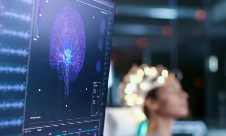
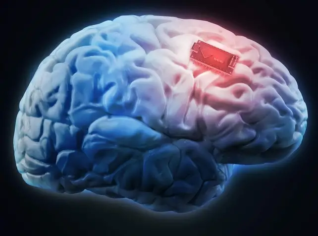
Technology and biology are blending together more and more. In live tissue, electrodes have been successfully generated by researchers at the Swedish universities of Linköping, Lund, and Gothenburg by using body molecules as triggers. The outcome, which was reported in the journal Science, paves the path for fully integrated electrical circuits to develop in living things. “We have been attempting to develop biologically inspired electronics for a number of decades to aid the growth of electrodes of the brain in the cranium. Now that we’ve let biology design our electronics, “explains Professor Magnus Berggren of Linköping University’s Laboratory for Organic Electronics (LOE).Understanding intricate biological processes, battling brain illnesses, and creating future machine-human interactions all depend on linking electronics to biological tissue. Conventional bioelectronics, on the other hand, have a fixed and static design that makes it challenging, if not impossible, to integrate with living biological signal systems. These devices were created concurrently with the semiconductor industry.

Researchers have created a technique for producing soft, substrate-free, electronically conductive materials in living tissue to close this biology-technology divide. By injecting a gel containing enzymes as the “assembly molecules,” the researchers were able to generate electrodes in the tissue of zebrafish and medicinal leeches. “The gel’s structure is altered by contact with bodily substances, which also causes it to become electrically conductive after injection. To start the electrical process, we can also change the gel’s composition based on the tissue “As one of the study’s principal authors and a researcher at LOE and Lund University, Xenofon Strakosas has said. To bridge this biology-technology gap, researchers have developed a method for generating soft, substrate-free, electronically conductive materials in living tissue. By injecting a gel containing enzymes as the “assembly molecules,” the researchers were able to construct electrodes in the tissue of zebrafish and medicinal leeches. “Contact with physiological fluids causes the gel’s structure to change, which also makes it more electrically conductive following injection. We can also alter the gel’s composition in accordance with the tissue to initiate the electrical process “Xenofon Strakosas, a researcher at LOE and Lund University and one of the study’s primary authors, has stated.

Endogenous chemicals produced by the body are sufficient to cause electrode development. In contrast to other research, there is no requirement for genetic change or external signals like light or electrical energy. The Swedish researchers achieved this first in the globe. Their research opens the door for a brand-new approach to bioelectronics. In the future, a viscous gel injection will suffice in place of implanted physical devices, which were previously required to initiate electronic operations in the body .Researchers also demonstrate in their paper that the technique may direct the electronically conducting material to particular biological substructures, resulting in the creation of functional interfaces for nerve stimulation. In the future, it might be possible to create fully integrated electronic circuits inside of biological things. The researchers successfully formed electrodes in zebrafish brain, heart, and tail fins as well as around the nerve tissue of therapeutic leeches during trials at Lund University. The gel injection had no negative effects on the animals, and the electrode generation had no negative effects either. Considering the animals’ immune systems was one of the studies’ numerous difficulties.
We created electrodes that were accepted by the immune system and brain tissue by cleverly altering the chemistry. Professor Roger Olsson spearheaded the investigation after learning about the electronic rose created by Linköping University researchers in 2015. The variation in cell structure between plants and mammals was one area of study concern. Animal cells resemble a mushy mass more than plant cells, which have hard cell walls that allow for the development of electrodes. Our findings provide for whole fresh perspectives on biology and electronics. Although there are still many issues to be resolved, this study is an excellent place to start “according to one of the major authors and LOE PhD student Hanne Biesmans.
Conclusion
These findings strongly suggest that either the installation of chronic electrodes or multiple recording electrodes during DBS does not raise the risk of brain hemorrhage or other intracranial problems, nor does it result in any biochemically identifiable harm to brain tissue.
let us know about what you think on this latest research? do you have anyne ith neurological desease? or ny